Tendon Diagram Labeled : Musculoskeletal System Skeletal Muscle Muscular System Anatomy Skeletal Muscle Anatomy. Related posts of muscles and tendons of the leg muscle anatomy head. Human muscle system, the muscles of the human body that work the skeletal system, that are under voluntary control, and that are concerned with movement, posture, and balance.broadly considered, human muscle—like the muscles of all vertebrates—is often divided into striated muscle (or skeletal muscle), smooth muscle, and cardiac muscle.smooth muscle is under involuntary control and is. To understand one of the most complex joints of our body i.e. The knee joint, you need a perfectly labeled diagram of the knee. The knee joins the thigh bone (femur) to the shin bone (tibia).
A labeled diagram of the knee with an insight into its working. Visceral muscle is found inside of organs like the stomach, intestines, and blood vessels. Human muscle system, the muscles of the human body that work the skeletal system, that are under voluntary control, and that are concerned with movement, posture, and balance.broadly considered, human muscle—like the muscles of all vertebrates—is often divided into striated muscle (or skeletal muscle), smooth muscle, and cardiac muscle.smooth muscle is under involuntary control and is. This diagram depicts human anatomy tendons and ligaments.human anatomy diagrams show internal organs, cells, systems, conditions, symptoms and sickness information and/or tips for healthy living. Tendon diagram muscle tendon diagram 9 out of 10 based on 20 ratings.
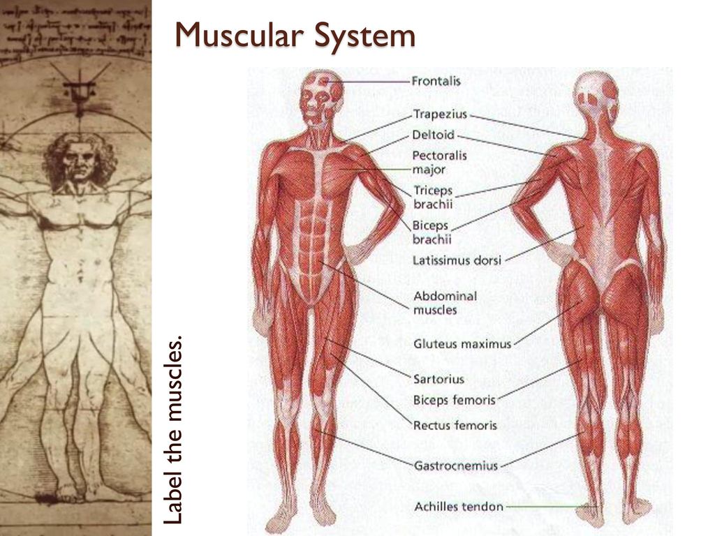
33 Muscular System Quiz Label Label Design Ideas 2020 from 9fc6ff.medialib.edu.glogster.com This muscular system chart shows in detail the deep layers of muscle on the back side of your body. More specifically, this beautifully illustrated anatomy chart includes neck and shoulders, multiple views of the back and spine, and frontal views of each muscular extremity of the human body. Jul 05, 2018 · the foot diagram has a complex structure made up of bones, ligaments, muscles, and tendons. If you want to manage a knee injury, you might have a good piece of knowledge on the dog knee anatomy.here, i will show you everything on the dog knee, including the bone involvement, ligaments, tendons and their arrangement with a labeled diagram. Each foot is made up of 26 bones, 33 joints and more than 100 muscles, tendons and ligaments, all of which work together to provide support,. Related posts of muscles and tendons of the leg muscle anatomy head. The knee joint, you need a perfectly labeled diagram of the knee. The patient would not notice much weakness in the upper limb due to the.
Read formulas, definitions, laws from muscle movements here.
The knee is one of the largest and most complex joints in the body. A muscle's origin is where a tendon attaches it to the *less* movable bone. This diagram depicts knee diagram tendons. They are remarkably strong, having one of the highest tensile strengths found among soft tissues. Use the location, shape and surrounding structures to help you memorize each muscle. This diagram depicts muscle in the body 744×1054 with parts and labels. However, the long head of the biceps brachii is one of the more common tendons to rupture. Shoulder tendons chart ~ labeled anatomy chart of shoulder ligaments on white background stocktrek images.a tendon is a structure that connects muscle to bone, and the biceps are connected by tendons at both the elbow and shoulder joints. The knee joins the thigh bone (femur) to the shin bone (tibia). Learn about the anatomy and physiology of tendons. The fleshy, thick part of the muscle is called its belly. The weakest of all muscle tissues, visceral muscle makes organs contract to move substances through the organ. Each foot is made up of 26 bones, 33 joints and more than 100 muscles, tendons and ligaments, all of which work together to provide support,.
Tendon diagram muscle tendon diagram 9 out of 10 based on 20 ratings. There are three types of muscle tissue: Read formulas, definitions, laws from muscle movements here. Labeled diagram view the muscles of the upper and lower extremity in the diagrams below. Branches of the femoral artery supply.
Muscles Of The Lower Leg And Foot Human Anatomy And Physiology Lab Bsb 141 from s3-us-west-2.amazonaws.com The patellar tendon attaches the patella to the top of the tibia. Learn about the anatomy and physiology of tendons. Make writing personal training programs easy with these custom designed exercise templates, and keep your clients focused and progressing. Leg muscle anatomy the legs are the lower limbs of the human body that provide support and stability in addition to allowing movement. Bones in shoulder, ligaments of the shoulder joint, parts of the shoulder joint, shoulder anatomy, shoulder joints and muscles, shoulder structure anatomy, shoulder tendon anatomy, shoulder tendons ligaments, human muscles, bones in shoulder, ligaments of the shoulder joint, parts of. This diagram depicts muscle in the body 744×1054 with parts and labels. The tendons have 2 functions: Muscular system anatomy muscle types.
In human anatomy, the peroneus longus (also known as fibularis longus) is a superficial muscle in the lateral compartment of the leg, and acts to evert and plantarflex the ankle.
The patellar tendon attaches the patella to the top of the tibia. A tendon is a structure that connects muscle to bone, and the biceps are connected by tendons at both the elbow and shoulder joints. A muscle's origin is where a tendon attaches it to the *less* movable bone. Tendon diagram ankle tendon diagram 9 out of 10 based on 90 ratings. In human anatomy, the peroneus longus (also known as fibularis longus) is a superficial muscle in the lateral compartment of the leg, and acts to evert and plantarflex the ankle. This will help you to understand the mechanism as well as the working. The tendons have 2 functions: Shoulder tendons chart ~ labeled anatomy chart of shoulder ligaments on white background stocktrek images.a tendon is a structure that connects muscle to bone, and the biceps are connected by tendons at both the elbow and shoulder joints. Each foot is made up of 26 bones, 33 joints and more than 100 muscles, tendons and ligaments, all of which work together to provide support,. Use the location, shape and surrounding structures to help you memorize each muscle. This diagram depicts muscle in the body 744×1054 with parts and labels. Tendon, tissue that attaches a muscle to other body parts, usually bones. 17 photos of the diagram of shoulder muscles and tendons.
Each foot is made up of 26 bones, 33 joints and more than 100 muscles, tendons and ligaments, all of which work together to provide support,. This diagram depicts human anatomy tendons and ligaments.human anatomy diagrams show internal organs, cells, systems, conditions, symptoms and sickness information and/or tips for healthy living. However, the long head of the biceps brachii is one of the more common tendons to rupture. The majority of muscles in the leg are considered long muscles, in that they stretch great distances. Link to pt program exercise templates.
35 Muscular System With Label Labels Database 2020 from slideplayer.com Muscle anatomy head 12 photos of the muscle anatomy head dog head muscle anatomy, human. In many cases, torn tendons begin by fraying. Tendon, tissue that attaches a muscle to other body parts, usually bones. This diagram depicts human anatomy tendons and ligaments.human anatomy diagrams show internal organs, cells, systems, conditions, symptoms and sickness information and/or tips for healthy living. Each foot is made up of 26 bones, 33 joints and more than 100 muscles, tendons and ligaments, all of which work together to provide support,. Labeled anatomy chart of shoulder ligaments on white background stocktrek images. If the complete motor neuron innervation to a muscle is lost. Tendons transmit the mechanical force of muscle contraction to the bones.
The only bone in the thigh is the femur, which extends from the hip to the knee.it can resist forces of 1,800 to 2,500 pounds, so it is not easily fractured.
Labeled anatomy chart with two bones, articular cartilage, joint cavity, synovial fluid, muscle. The weakest of all muscle tissues, visceral muscle makes organs contract to move substances through the organ. The knee joins the thigh bone (femur) to the shin bone (tibia). Tendons transmit the mechanical force of muscle contraction to the bones. A tendon is a structure that connects muscle to bone, and the biceps are connected by tendons at both the elbow and shoulder joints. The dog knee injury is very common in the field. Jul 05, 2018 · the foot diagram has a complex structure made up of bones, ligaments, muscles, and tendons. In the back and elsewhere in the body, tendons attach muscles to bones. Make writing personal training programs easy with these custom designed exercise templates, and keep your clients focused and progressing. The legs include the upper leg, knee, lower leg, ankle, and foot. This muscular system chart shows in detail the deep layers of muscle on the back side of your body. Visceral muscle is found inside of organs like the stomach, intestines, and blood vessels. On the other hand, the insertion is where a tendon attaches that muscle to the *more* movable bone.


0 Comments:
Posting Komentar